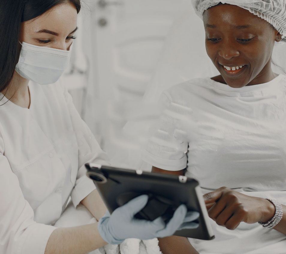
Surgical
Dermatology
-
How?
Surgical excision is recommended for malignant (cancerous) and benign (non-cancerous) skin lesions and growths.
Read More -
What?
This procedure is performed under local anesthetic in our fully-equipped office and takes approximately 30 minutes, depending on the size of the lesion. Once the target lesion is removed, it is sent to the lab for microscopic examination, and the area is brought back together with stitches to allow a faster healing time. These stitches may be dissolvable, where they will be absorbed by your body over the next 90 days, or non-dissolvable, where they will need to be removed in 7-14 days. Depending on the treatment area, it takes approximately 2-6 weeks to heal from an excisional procedure, however, most can immediately resume normal daily activities.
Read More -
Why?
We always recommended follow-up appointments after excisional procedures to address and optimize the cosmetic results of the scar.
Read More
-
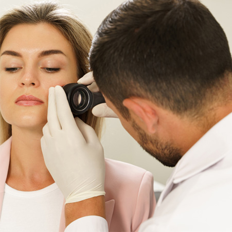
Basal Cell Carcinoma (BBC)
Basal cell carcinoma is the most common type of skin cancer. It appears as a small red, pink pimple that will continue to grow. It has various patterns of growth and can spontaneously bleed. It can be effectively treated with many different options including liquid nitrogen, excision, Mohs Micrographic Surgery, and superficial radiation treatment.
Read More -
Squamous Cell Carcinoma (SCC)
Squamous cell carcinoma( SCC) is a type of skin cancer that appears as a scaly red/pink spot that does not appear to go away. These can grow quickly and spread if not detected early. SCC tends to occur in those who have had years of sun exposure and/or tanning bed use over an extended period of time. It can be especially dangerous in patients who are immunosuppressed. Early detection can be treated with Liquid nitrogen, excision, Mohs Micrographic surgery, and superficial radiation treatment.
Read More -
Malignant MelanomaRead More
Malignant melanomas are a type of skin cancer that is derived from the pigmented melanocytes in your skin. It can affect a patient of any skin color and can be metastatic if left alone for several years. Early detection is very important in the treatment and cure rate for this type of skin cancer.
There are 5 key aspects to look for when looking for a melanoma or dysplastic growth:
- A - Asymmetry, appears when one side of the lesion does not appear like the other.
- B - Border becoming irregular
- C - Color, various colors noted within the lesion
- D - Diameter, the growth of the lesion is greater than 6 mm in size
- E - Evolution, the lesion growing, changing, or evolving.
Treatment for any type of melanoma is surgical intervention. These tumors have a great recovery rate when caught early and treated.
-
Actinic Keratosis
Actinic keratosis are pre-cancerous lesions that appear to be red/pink scaly lesions. Usually, these are easier to see than to treat and tend to develop due to chronic sun exposure. As a small percentage of these can develop into skin cancers, it is important to treat them early.
Read More

-
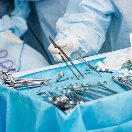
What is MOHS Surgery?
Developed by Frederic E. Mohs, M.D. in the 1930s, Mohs Micrographic Surgery is a highly precise, effective method that excises not only visible skin cancer but also any “roots” that may have extended beneath the surface of the skin. The five-year cure rates have been demonstrated up to 99 percent for first-treatment cancers and 95 percent for recurrent cancers.
Read More -
Why?
Mohs surgery is most commonly used for basal and squamous cell carcinomas, although it can be recommended for the eradication of other cancers such as melanoma. Cancers that are likely to recur or have already recurred are often treated using this technique because it is so thorough. High precision makes Mohs surgery ideal for the elimination of cancers in cosmetically and functionally critical areas.
Read More -
How?
Mohs surgery involves the systematic removal and microscopic analysis of thin layers of tissue at the tumor site until cancer has been completely eliminated. The immediate and complete microscopic examination and evaluation of excised tissue are what differentiates Mohs surgery from other cancer removal procedures. The only cancerous tissue is removed, minimizing both post-operative wound size and complete removal of the cancer is confirmed microscopically reducing the chance of recurrence. In addition, the entire tumor is cleared and the site repaired in one session. Mohs physicians are highly trained to function as surgeons, pathologists, and reconstructive surgeons during the cancer removal process.
Read More